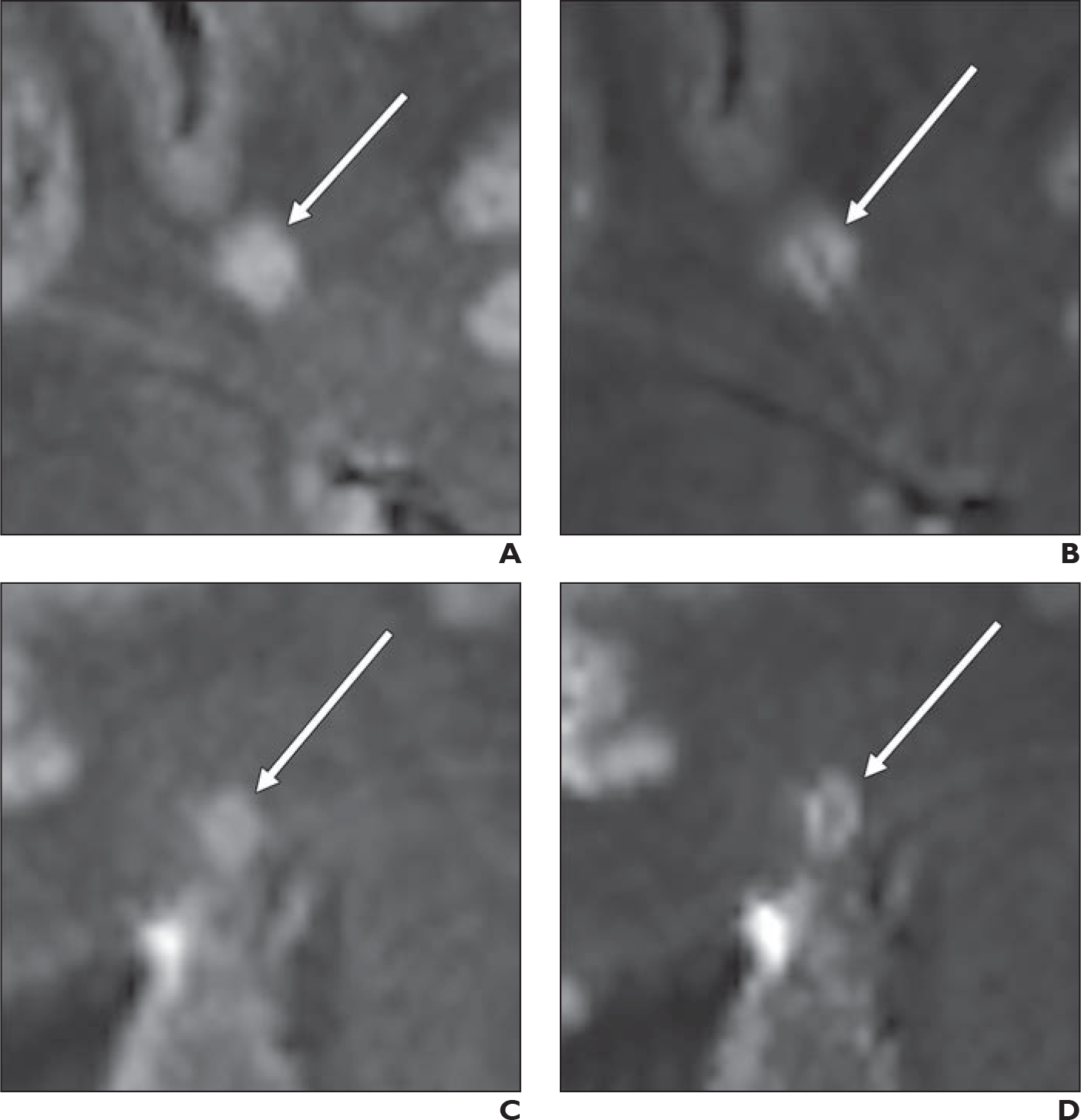
Fig. 4—

Comparison of precontrast and postcontrast FLAIR* images in terms of central vein sign (CVS) visualization in white matter lesions (WMLs). In this example, patient is 43-year-old participant with multiple sclerosis.
A and B, WML (arrow) was classified as CVS-negative on precontrast image (A) but as CVS-positive on postcontrast image (B).
C and D, WML (arrow) was classified as CVS-negative on precontrast image (C) but as CVS-positive on postcontrast image (D).
