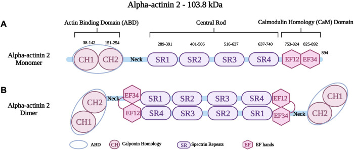
FIGURE 2.
(A) Schematic representation of alpha-actinin two monomer with different residues in the Actin Binding Domain (ABD), Spectrin repeats (SR) and EF hands (EF). (B) Schematic representation of alpha-actinin two dimer showing the interaction between the neck region and the EF hands. Created with BioRender.com. Adapted from (Haywood et al., 2016).

