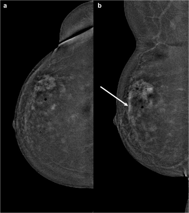
Fig. 3.

CESM images of a 56-year-old woman with histopathological proven breast cancer in the upper outer quadrant of the right breast. Images were acquired with machine (b). Although the malignancy can be clearly detected, the diffuse surrounding enhancement makes the detection of possible satellite lesions and a possible extension in the direction of the mammilla (arrow) almost impossible. The images were rated as showing a marked background enhancement (4/4)
