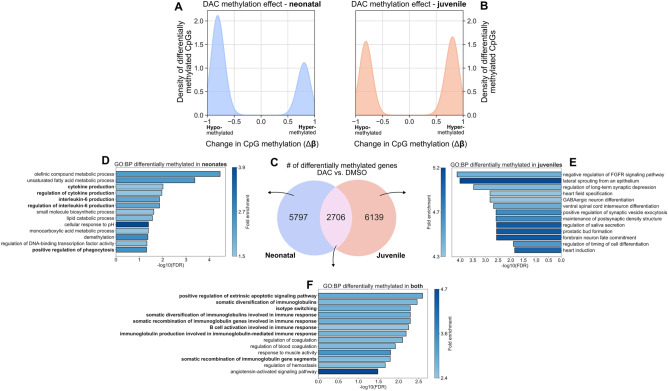
Figure 2.
DAC treatment of mice infected with E. coli induces CpG demethylation at unique genomic loci and with greater strength in neonates versus juveniles. (A, B) Density plot of changes in CpG methylation frequency (Δβ) in (A) neonatal mice and (B) juvenile mice. DAC treatment more strongly induces demethylation (Δβ < 0) in neonates than in juveniles, which are less likely to be demethylated (Δβ > 0). (C) Venn diagram showing the number and overlap of differentially methylated genes with at least one differentially methylated CpG in DAC vs. DMSO across neonates and juveniles. (D–F) Over-representation analysis (ORA) from gProfiler showing GO: Biological Processes (GO:BPs) associated with genes differentially methylated in response to DAC treatment in (D) neonates, (E) juveniles, and (F) both.

