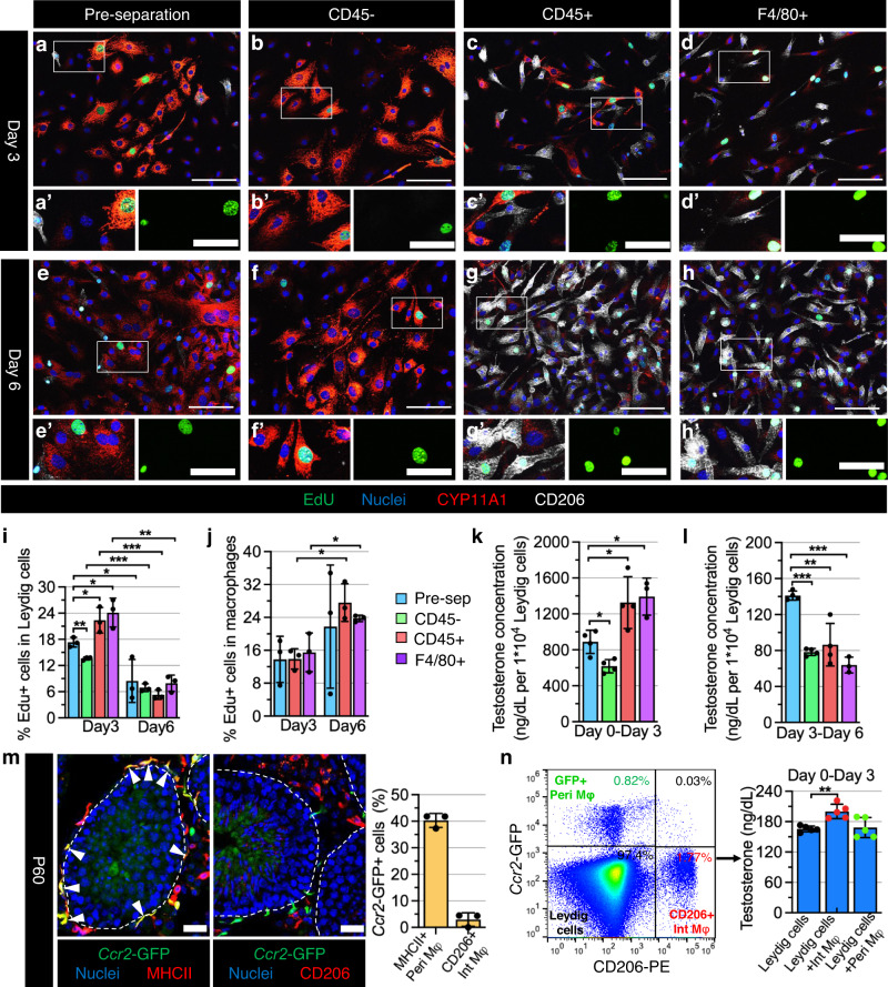
Fig. 7. Adult interstitial macrophages promote Leydig cell proliferation and steroidogenesis.
a–h Representative images (n = 3 independent experiments) of primary cell culture after 3 days (a–d) and 6 days (e–h) from adult (3-month-old) C57BL/6 J testes for pre-separation (a, e), CD45-depleted (b, f), CD45-enriched (c, g), and F4/80-enriched (d, h) populations. i–l Graphs showing quantification (n = 3 independent experiments from 6 testes) of percent EdU+ Leydig cells (i), percent EdU+ macrophages (j), and testosterone concentration (n = 4 independent experiments from 8 testes) in culture media after 0–3 days of culture (k) and 3–6 days of culture (l) in the 4 different cell populations. m Representative images of Ccr2GFP/+ testes and graph showing quantification (n = 3 independent testes) of percent GFP-expressing interstitial (CD206+) and peritubular (MHCII+) macrophages at P60. Arrowheads denote GFP-expressing MHCII+ peritubular macrophages. n FACS isolation and culture of testicular macrophages from adult Ccr2GFP/+ testes. Flow cytometry plot (left) shows peritubular macrophages (Peri Mφ: GFP+CD206–), interstitial macrophages (Int Mφ: GFP–CD206+) and Leydig cells (GFP–CD206–) gated on total live cells as indicated; graph (right) shows testosterone concentrations (n = 5 independent experiments from 10 testes) in culture media after 3 days of culture of Leydig cells alone and Leydig cells co-cultured with Int Mφ or Peri Mφ. Thin scale bar, 100 μm; thick scale bar, 25 μm. All graph data are shown as mean +/– SD. *P < 0.05; **P < 0.01; ***P < 0.001 (two-tailed Student’s t test). Exact P values are provided in the Source Data file.

