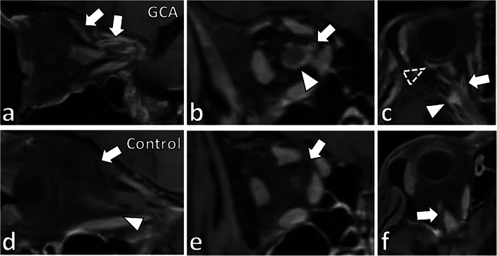
Fig. 2.
Vessel wall enhancement of the ophthalmic artery and its branches in a patient diagnosed with GCA. In a patient diagnosed with GCA, (a) sagittal, (b) coronal, and (c) axial BB-MRI images show concentric vessel wall thickening and enhancement of the right ophthalmic artery (a–c, arrows). There was also optic nerve sheath enhancement (b–c, arrowheads) and partial rim enhancement posterior to the globe (c, dashed arrowhead). In an age-matched control case, (d) sagittal, (e) coronal, and (f) axial BB-MRI show no vessel wall thickening or enhancement of the ophthalmic artery (d–f, arrows). No pathologic enhancement of the optic nerve sheath (d, arrowhead) or other orbital structures was identified

