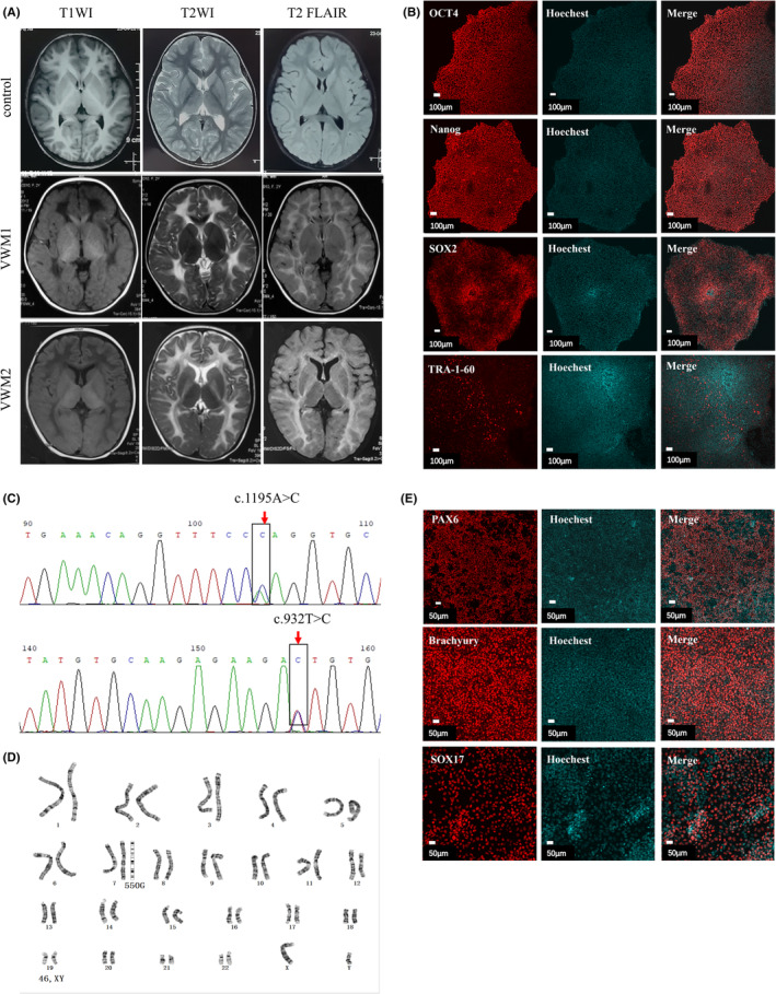
FIGURE 1.

VWM patient‐derived iPSC and identification. (A) Brain MRI of one normal control child (3 years old) and two children with VWM. VWM1 and VWM2 are cases of childhood onset, and the age of onset and MRI age are 12 months and 3 years old, respectively. The white matter of the lesions in children with VWM showed low signal on T1WI, high signal on T2WI, and low signal on T2 FLAIR part (liquefaction sign). The earlier the onset, the more extensive the white matter involvement. (B) Immunofluorescence analysis of pluripotent markers, OCT4, Nanog, SOX2, and TRA‐1‐60, scale bar 100 μm. (C) Sanger generation sequencing analysis of the genomic DNA from VWM2 patient‐derived iPSC showed: EIF2B4 c.1195A>C; c.932T>C. (D) Karyotype analysis showed 46, XY. (E) Immunofluorescence staining of differential marker: endoderm (SOX17), mesoderm (Brachyury), and ectoderm (PAX6), scale bar 50 μm.
