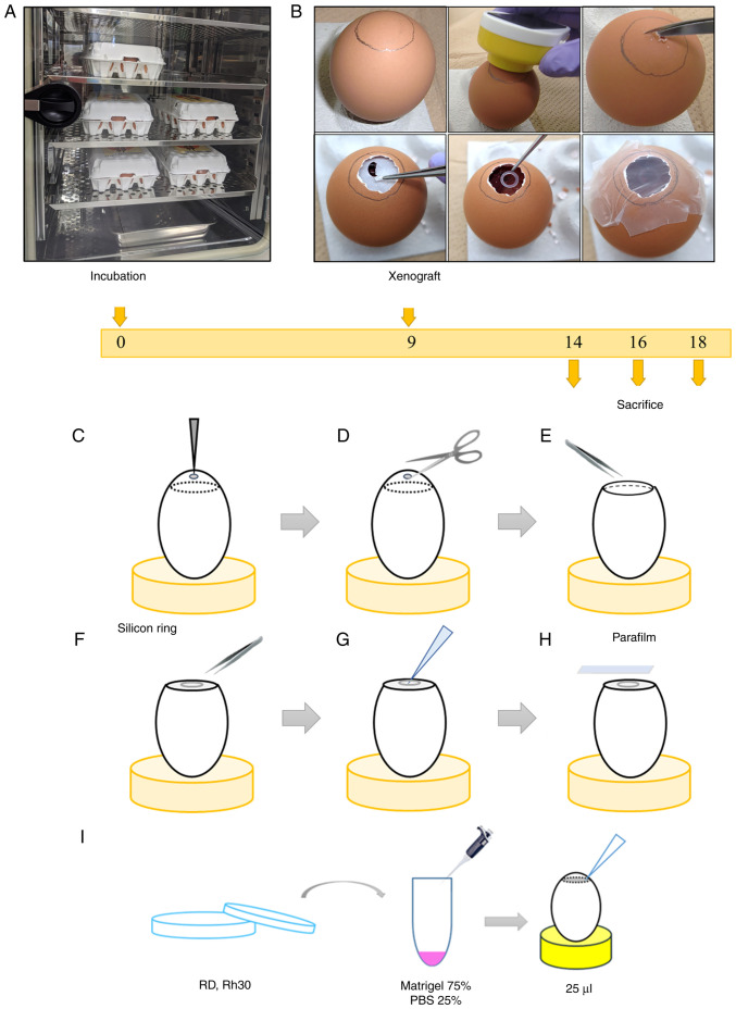
Figure 1.
Diagram of cell-derived xenograft model generation using the CAM assay. (A) Day 0. Commercial fertilized eggs were placed upright in an incubator at 37°C and 60% humidity. (B) Day 9. Photographs of steps (C-H). Prior to step C, the edges of the air chamber were traced with a pencil while illuminating the eggs in the darkroom. (C) A hole was made on top of the egg with an egg piercer. (D) Scissors were used to cut the shell along the edge of the air chamber. (E) Eggshell membrane was removed with tweezers. (F) A silicon ring was placed on the CAM. (G) A cell suspension (25 µl) was grafted onto the CAM. (H) After cell inoculation, the window in the egg was tightly sealed using parafilm and the eggs were returned to the incubator. (I) Cell suspension was prepared by detaching tumor cells from culture dishes using trypsin/ethylenediaminetetraacetic acid and counting them. The cells were resuspended in Matrigel and PBS (3:1 ratio) at 2.0×10⁶ cells/25 µl Matrigel solution. CAM, chorioallantoic membrane; PBS, phosphate-buffered saline.

