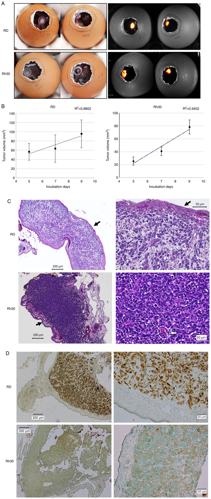
Figure 2.
Establishment of a cell-derived xenograft model on the CAM using the RMS cell lines, RD and Rh30. (A) Tumor formed on the CAM on day 16 (7 days after transplantation of RD or Rh30 cells). Images on the left were captured on a clean bench, whereas images on the right were observed using the G:BOX Chemi XRQ gel doc system following the addition of luciferin. (B) Temporal changes in tumor volume. Tumors were resected on days 14, 16 and 18, and the volume was calculated using Vernier caliper measurements. (C) Hematoxylin and eosin staining of the resected tumors on day 16 (left, ×40 magnification; right, ×200 magnification). Accumulation of cells and formation of RMS tissue along the CAM (black arrow), and infiltration of some chick red blood cells into the tissue (inside white dotted line) were observed. (D) Immunohistochemical staining of anti-human vimentin in the tumor tissue (left, ×40 magnification; right, ×200 magnification). Counterstaining of sections was performed with methyl green. These results indicated that the resected tumor consisted of human RMS cells transplanted on day 9. CAM, chorioallantoic membrane; RMS, rhabdomyosarcoma.

