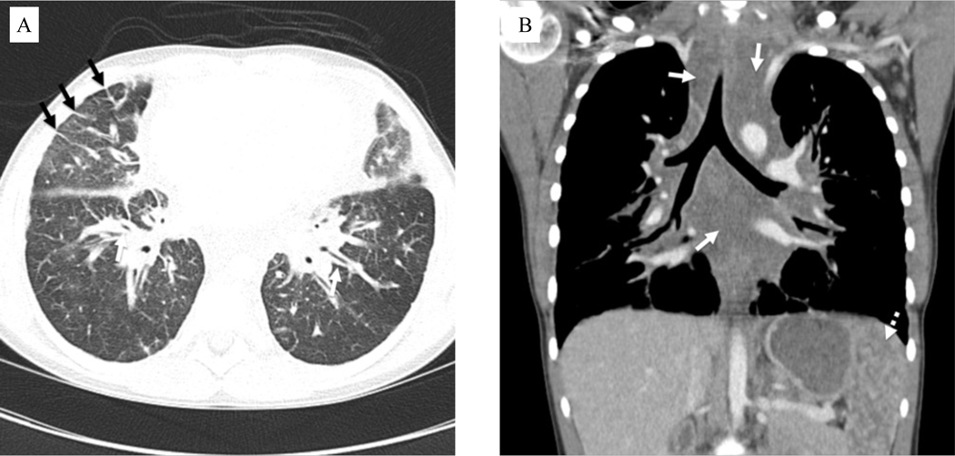
Figure 2.
Computed tomography images of patient with kaposiform lymphangiomatosis. A) Axial contrast-enhanced chest image in lung windows in an 11-year-old female shows variable degrees of interstitial thickening, visible along the bronchial walls centrally (white arrows) and secondary pulmonary lobules peripherally (black arrows). B) Coronal contrast-enhanced chest image in the same patient shows expansion of the mediastinum by poorly defined fluid attenuation tissue (solid white arrows), corresponding to infiltration by disease. Numerous lesions are also noted in the spleen (dotted white arrow).

