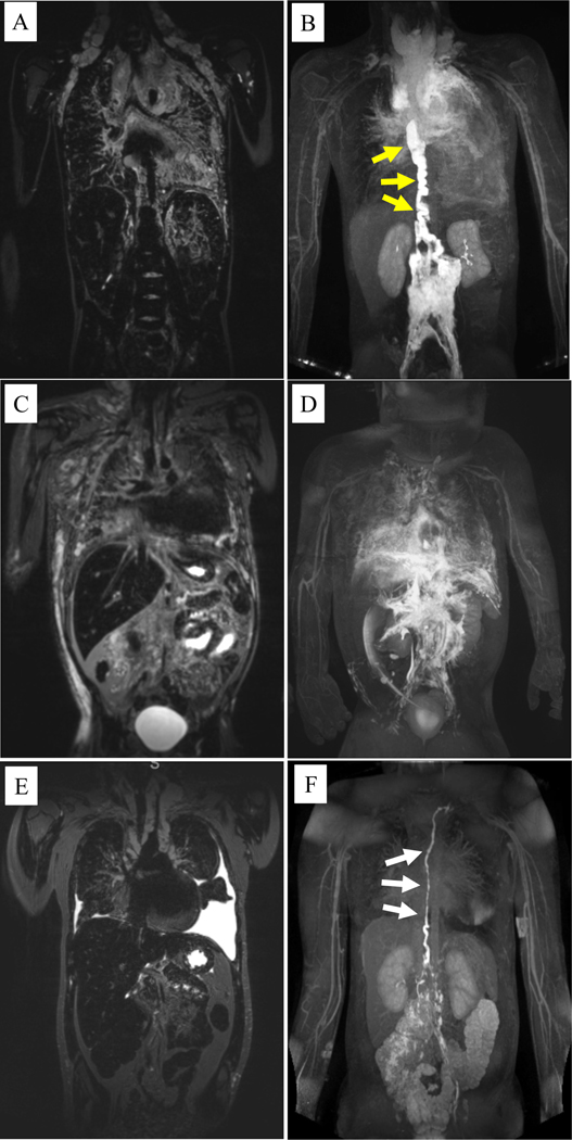
Figure 4.
Coronal T2-weighted magnetic resonance imaging (MRI) and magnetic resonance lymphangiography (MRL) of patients with kaposiform lymphangiomatosis. A) T2-weighted MRI of a 10 year-old male shows significant mediastinal thickening with pulmonary edema and bilateral supraclavicular edema. B) The corresponding MRL shows a significantly dilated and tortuous thoracic duct (yellow arrows) that terminates in the mediastinum with significant perfusion of the mediastinal and pulmonary lymphatics on MRL. C) T2-weighted MRI of a 4 year-old male shows mediastinal thickening with pulmonary edema, dermal edema, and ascites. D) The corresponding MRL shows no clear thoracic duct but several abnormal lymphatic channels coursing into the thorax leading to mediastinal and pulmonary lymphatic perfusion. E) T2-weighted MRI of a 12 year-old female shows mediastinal thickening with bilateral pleural effusions. F) The corresponding MRL shows a dilated and mildly tortuous thoracic duct (white arrows), but with limited mediastinal or pulmonary lymphatic perfusion.

