Fig. 6.
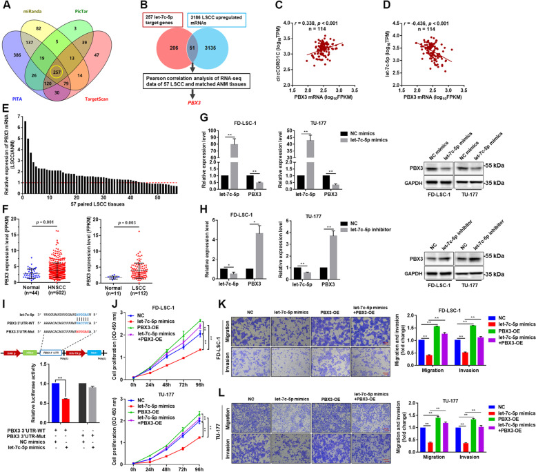
PBX3 is a direct target gene of let-7c-5p, which acted as an oncogene in LSCC cells. a Venn analysis of the target genes of let-7c-5p predicted by miRanda, PicTar, PITA, and TargetScan. b Integrated analysis of bioinformatics-predicted target genes and RNA sequencing data of 57 pairs of LSCC tissues was performed to screen for let-7c-5p target genes. c & d Correlation analysis between circCORO1C (c) or let-7c-5p (d) and PBX3 expression using RNA sequencing data of 57 pairs of LSCC tissues and matched ANM tissues. e PBX3 expression in RNA sequencing data of 57 pairs of LSCC tissues and matched ANM tissues. The expression levels of PBX3 in each LSCC tissue were normalized to corresponding matched ANM tissue. f Analysis of PBX3 expression in HNSCC and LSCC tissues using transcriptome sequencing data from TCGA database. g & h FD-LSC-1 and TU-177 cells were transfected with let-7c-5p mimics (g), let-7c-5p inhibitor (h) or NC, and PBX3 expression was detected by qPCR and western blotting. i HEK293T cells were co-transfected with let-7c-5p mimics and wild-type or mutant PBX3 3′ UTR reporter plasmids, and luciferase reporter assays were performed to evaluate the effect of let-7c-5p on luciferase activity. j FD-LSC-1 and TU-177 cells were transfected with let-7c-5p mimics or co-transfected with let-7c-5p mimics and PBX3 overexpression plasmids, and CCK8 assay was performed to detect cell proliferation. k & l FD-LSC-1 (k) and TU-177 (l) cells were transfected with let-7c-5p mimics or co-transfected with let-7c-5p mimics and PBX3 overexpression plasmids. Changes in cell migration and invasion capacity were evaluated by Transwell assays. Data are presented as the means ± SD of three independent experiments. *P < 0.05; **P < 0.001

