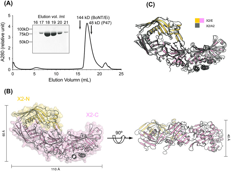
Figure 4. Crystal structure of BoNT/E OrfX2.
(A) SEC analysis of the recombinant OrfX2 (n=3, a representative result is shown). Purity of the peak fractions is examined by SDS-PAGE. The peak positions of two reference proteins with molecular weights of 144 kDa and 48 kDa are indicated by arrows. (B) Ribbon and surface representations of OrfX2 in two different views. The OrfX2-N domain is colored yellow and the OrfX2-C domain in pink. (C) Structure alignment of OrfX2 of BoNT/E (shown in yellow and pink) and a previously reported OrfX2 of BoNT/A2 (PDB: 6EKV, shown in grey).

