FIGURE 4.

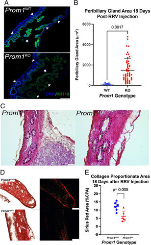
Prom1 null mutation results in dilated PBGs 18 days after RRV injury compared with Prom1-expressing PBGs. (A) Mice homozygous for the Prom Cre/Cre allele were bred, resulting in functional Prom1 knockout (Prom1 KO) mice. (A) Fluorescence imaging of KRT19 expression in both WT and Prom1 KO EHBD. Triangles point to PBGs, and stars signify EHBD lumen. Scale bar, 250 μm. (B) PBGs in Prom1 KO are larger than WT (n=3 biological replicates). (C) Hematoxylin and eosin staining of PBGs in the EHBD demonstrates enlarged PBGs in Prom1 KO as compared with Prom1 WT. PBGs are outlined with dashed lines. Scale bars, 50 μm. (D) Sirius red staining of EHBD from Prom1-expressing (Prom1 Cre/+) and Prom1 KO 18 days after RRV injury. Scale bar, 100 μm. (E) Prom1-expressing EHBD (Prom1 Cre/+) CPA is statistically significantly higher than Prom1 KO EHBD (p < 0.005). Abbreviations: CPA, collagen proportionate area; EHBD, extrahepatic bile duct; PBG, peribiliary gland; Prom1, Prominin-1; RRV, rhesus rotavirus; WT, wild-type.
