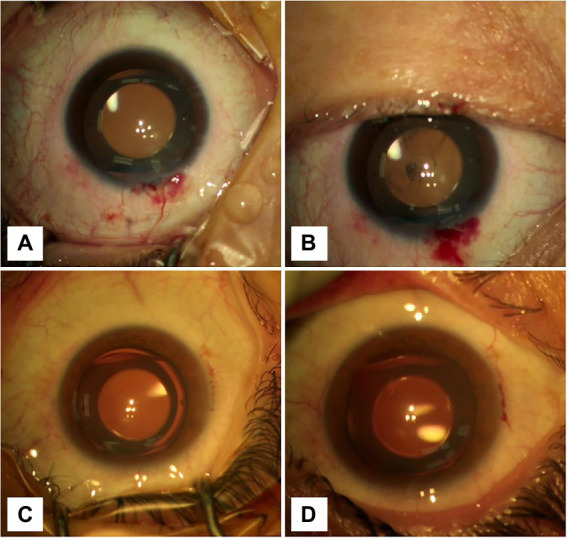
Figure 2.

Graphs showing the changes in the anterior chamber before and after removing the blepharostat. (A) The CCI was located at 12 o’clock. The anterior chamber was clean, and the incision was well self-sealed before blepharospasm removal. (B) The CCI was located at 12 o’clock. After removing the blepharospasm, the conjunctival sac hemorrhagic fluid flowed into the anterior chamber. (C) The CCI was located at 9 o’clock. The anterior chamber was clean, and the incision was well self-sealed before blepharospasm removal. (D) The CCI was located at 9 o’clock. After removing the blepharospasm, any visible inflow of orbital surface fluid was not seen.
