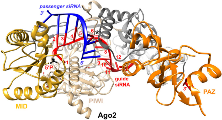
FIGURE 8.
View of the crystal structure of Ago2 bound to an RNA duplex (PDB ID 4W5T) kinked between positions 6 and 7 of the antisense (guide) strand (asterisk). Ago2 domains are highlighted and labeled, and the siRNA (antisense and sense) strands are colored in red and blue, respectively. Antisense strand residues 1 to 12 are numbered, and the 5′-phosphate group is highlighted in black and ball-and-stick mode.

