Fig. 3.
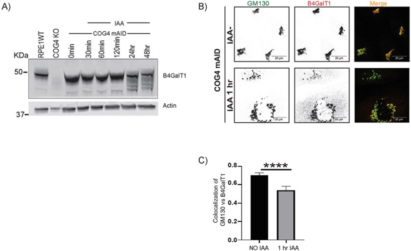
Acute depletion of COG4-mAID-mCherry displaces the B4GalT1 enzyme from the Golgi into CCD vesicles. (A) The hTERT RPE1 COG4 KO/OsTir1-9myc/hCOG4-mAid-mCherry cells were treated with 0.5 mM IAA for different times as indicated. Cells were collected and lysed, and the expression of B4GalT1 and actin was analyzed by WB. No depletion of B4GalT1 was observed within 2 h of treatment, but the B4GalT1 band was more diffuse at 24 h IAA treatment, indicating protein degradation. (B) IF analysis of untreated and IAA treated cells. The COG4-mAID clone was treated 0.5 mM IAA for 1 h. The cells were fixed and stained for GM130 (green) and B4GalT1 (as red). Note that the acute depletion of COG4-mAID-mCherry displaced the enzyme from the Golgi into vesicle-like structures. Scale bars, 20 μm. (C) Quantification of colocalization between GM130 and B4GalT1 from n = 2 independent experiment with >30 cells analyzed. ****, P < 0.0001, significant. Error bar represents mean ± SD. The green and red channels are presented in inverted black and white mode, whereas the merged view is shown in RGB mode. Scale bars, 20 μm

