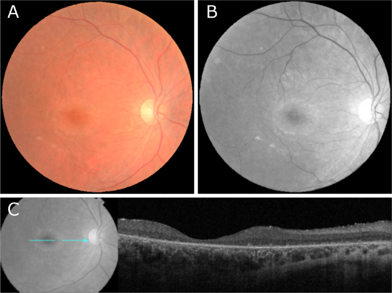
Figure 7.
Fundus photography and OCT performed in a patient with confirmed biallelic variants in GRN/CLN11. (A, B) Zeiss color and red-free autofluorescence imaging in patient XI revealed no abnormalities. (C) OCT imaging of patient XI revealed diffuse retinal thinning, loss of outer retinal structure, and loss of the ellipsoid zone.

