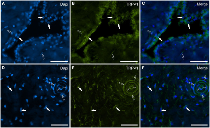
Figure 6.
Photomicrographs of the cryosections of the synovial membrane of a horse metacarpophalangeal joint showing transient receptor potential vanilloid 1 (TRPV1) (B, E), immunoreactivity in synoviocytes (A–C), and fibroblast and vascular cells (D–F). (A–C) The white arrows indicate the DAPI (Blue) labeled nuclei of some round synoviocytes expressing bright TRPV1 (Green) immunoreactivity. The open arrows indicate subintimal cells (likely fibroblasts) showing faint-to-moderate TRPV1 immunoreactivity. (D–F) The open arrows indicate the DAPI (Blue) labeled nuclei of some cells of the interstitial connective tissues of the synovial membrane (close to the subintima) expressing moderate-to-bright TRPV1 (Green) immunoreactivity. The open arrows and the small open arrows indicate the DAPI labeled nuclei of the vascular smooth muscle cells and endothelial cells, respectively, expressing moderate TRPV1 immunoreactivity. (C, F) Merged images. Scale bar = 50 μm.

