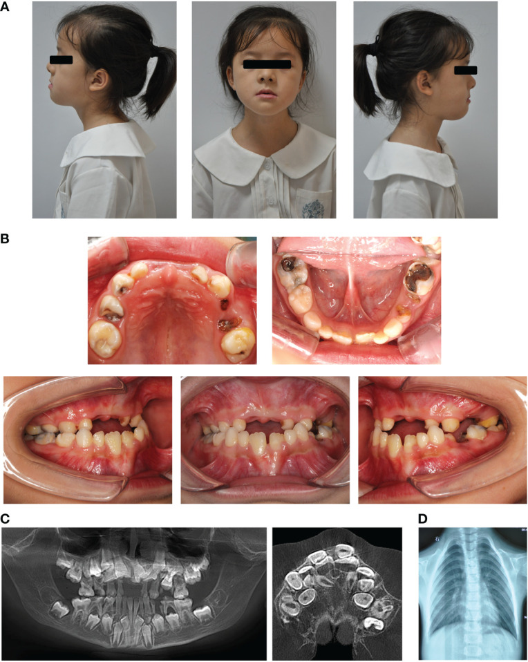
Figure 1.

Typical clinical and radiological findings in the proband with CCD. (A) Front and profile photographs of the individual showing a concave face, hypoplasia of the maxilla, and a depressed nasal bridge. (B) Intraoral images showing enamel and dentin hypoplasia. (C) Panoramic view showing embedded permanent, supernumerary, and retention of deciduous teeth. (D) Chest radiograph showing hypoplasia of the left clavicle.
