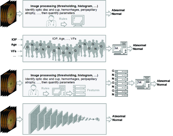
Figure 2.
Evolution of AI in glaucoma. First row: Image processing and expert systems were used to identify glaucoma landmarks or features (such as cup-to-disc ratio or hemorrhages) from retinal images with the assistance of a glaucoma specialist and glaucoma landmarks are identified. Second row: Numerical parameters like raw visual fields (VFs), intraocular pressure (IOP), and age from normal and glaucomatous subjects (presented as N and G) are input to a conventional machine learning model (e.g., neural network) without glaucoma specialist assistance and diagnosis is made. Third row: Image processing and expert systems were used to quantify glaucoma landmarks (extract features) with the assistance of a glaucoma specialist then quantified parameters (features) from normal and glaucomatous subjects are fed to a conventional machine learning model to make diagnosis. Fourth row: Retinal image is fed to an end-to-end deep learning model and the diagnosis is made without assistance from a glaucoma specialist.

