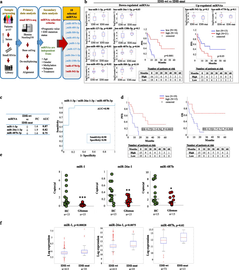
Fig. 1.
Genome-wide analysis and selection of a circulating miRNA-signature deregulated in glioma patients based on IDH-mutation. a Schematic representation of the experimental workflow for the global miRNA profiling by small RNA-seq; b) Box-plots and Kaplan-Meier curves based on the indicated miRNA expression levels in serum samples of glioma patients. In the box-plots serum miRNA levels in IDH-wild type (wt) vs. IDH-mutant (mut) patients are reported as Log2, boxes define the 25th and 75th percentiles, the horizontal line in the boxes indicates the median, and bars define the minimum and maximum values. The Kaplan-Meier plots indicate the relation between the miRNAs downregulated (left panel) or upregulated (right panel) in IDH-wt vs. IDH-mut patients and the PFS. Patients with high and low signal intensity for a specific miRNA signature were defined by considering positive and negative z-score values of the mean intensity values of the miRNAs. c Fold change expression (FC) and area under the ROC curve (AUC) of the indicated miRNAs and ROC curve plotted for diagnostic potential and discriminatory accuracy of serum miR-1/−26a-1/−487b combination. d Kaplan-Meier survival plots for PFS and OS based on miR-1/−26a-1/−487b expression levels. e Scatter plots of the expression levels of the indicated miRNAs, analysed by digital PCR, in serum samples of glioma patients vs. healthy controls (HC). f Box-plots of the indicated miRNA expression levels in IDH-wt vs. IDH-mut tumour tissue samples from a LGG dataset of the TCGA (left and middle panels) and from a GBM dataset of the CGGA (right panel); values are reported as Log2. Boxes define the 25th and 75th percentiles. The horizontal line in the boxes indicates the median and the edges of the box are the 25th and 75th percentiles * = p ≤ 0.05, ** = p ≤ 0.01, *** = p ≤ 0.001

