Fig. 6.
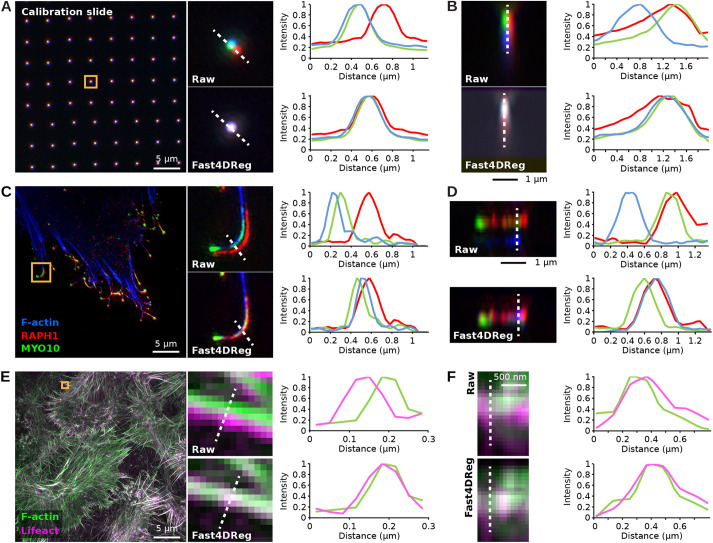
Fast4DReg can align 3D multichannel images. (A,B) A three-channel 3D calibration slide image was aligned using Fast4DReg. Merged images and line-intensity profiles (along the dashed white lines) are displayed to highlight the level of overlap between the three channels. (A) A z-projection is displayed to visualize the lateral misalignment corrected by Fast4DReg. (B) A y-projection of one of the calibration slide spots is displayed to illustrate the axial misalignment corrected by Fast4Dreg. (C,D) The drift table generated in A,B was then used to correct a 3D SIM image of a U2-OS cell expressing GFP-tagged lamellipodin (RAPH1, red) and MYO10–mScarlet (green), and labeled to visualize its actin cytoskeleton (blue). (C) A z-projection is displayed to visualize the lateral misalignment, evident in filopodia, corrected by Fast4Dreg. (D) A y-projection of one filopodium visualizes the axial misalignment corrected by Fast4Dreg. (E,F) A 3D SIM image of MCF10DCIS.com cells expressing RFP–Lifeact (magenta) and stained to visualize F-actin (green) was aligned using Fast4DReg directly. (E) A z-projection is displayed to visualize the lateral misalignment corrected by Fast4DReg. (F) A y-projection visualizes the slight axial misalignment corrected by Fast4DReg.

