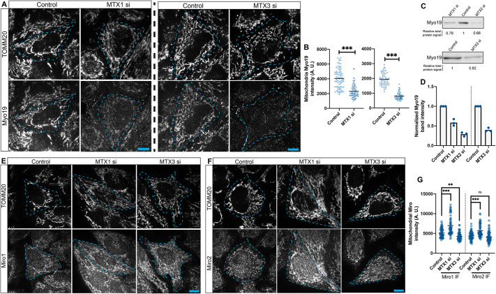
Fig. 3.
Metaxins are essential for Myo19 recruitment to mitochondria. (A) Immunofluorescence images of TOMM20 and Myo19 signals in cells treated with either a control oligonucleotide, or MTX1 or MTX3 siRNAs. (B) For these groups, quantitation of mitochondrial Myo19 intensity is shown. A two-tailed unpaired Student's t-test was used to determine significance. (C,D) Western blots (C) for Myo19 in cells treated with either a control oligonucleotide, or MTX1, MTX2 siRNA or MTX3 siRNAs, and quantitation of the bands (D). (E,F) Immunofluorescence images of either control, MTX1-depleted or MTX3-depleted cells, in which TOMM20 and Miro1 (E) or Miro2 (F) signals are shown. (G) Quantitation of mitochondrial Miro1 and Miro2 intensity in control cells, cells lacking MTX1 and cells lacking MTX3. One-way ANOVA and Dunnett's multiple comparisons test was used. Three independent experiments were performed; n=20–30 cells for each experiment. In graphs, the bars represent the means. In the images, cell boundaries are outlined. All scale bars: 10 μm. A.U., arbitrary units; IF, immunofluorescence. ns, not significant; **P<0.005; ***P<0.0005.

