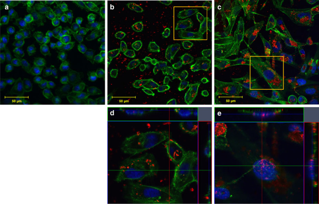
Fig. 8.
MDA-MB-231 cellular uptake and binding after a 0-min, b 60-min, and c 24-h incubation with SWNH–QD complexes. d, e Orthogonal snapshots of the regions of interest in b and c, respectively. The cross sections in the orthogonal images indicate SWNH–QDs within the nucleus and within the cytoplasm. Staining represented as: green Oregon Green® phalloidin F-actin stain; blue DAPI nuclear stain; and red SWNH–QD conjugates. (Color figure online)

