. 2023 Mar 17;12:e80768. doi: 10.7554/eLife.80768

Figure 7. Development of alopecia areata (AA) in the humanized mouse model treated with ILC1lc.

(A) Significant hair loss is observed following the injection of ILC1lc and enriched CD8/NKG2D cells, while in the PBMCs/PHA treated group, hair number remains almost constant. N=7–9 xenotransplants/group from three independent donors, Following Shapiro-Wilk test, Mann-Whitney U test: #p<0.05, ##p<0.01. (B) HF dystrophy and perifollicular lymphocytic infiltrates around anagen hair follicles (HFs) (H&E staining) combined with strong expression of (C) HLA-A,B,C, β2 MG, HLA-DR, and downregulation of α-MSH and TGF-β1 in the ILC1lc and in (D) enriched CD8/NKG2D cells versus xenotransplants treated with (E) PBMCs/PHA (IHC staining) (F) quantitative data. N=5–9 xenotransplats/group from three independent donors. 4–5 defined reference areas were evaluated per section, and three sections per xenotransplants. Following Shapiro-Wilk test, Student’s t-test: *p<0.05, **p<0.01, ***p<0.001. Scale bar, 50 µm. DP - dermal papilla, HM - hair matrix.

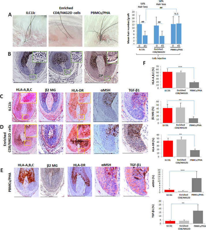
Figure 7—source data 1. Quantitative data for development of AA in the humanized mouse model treated with ILC1lc.

Figure 7.

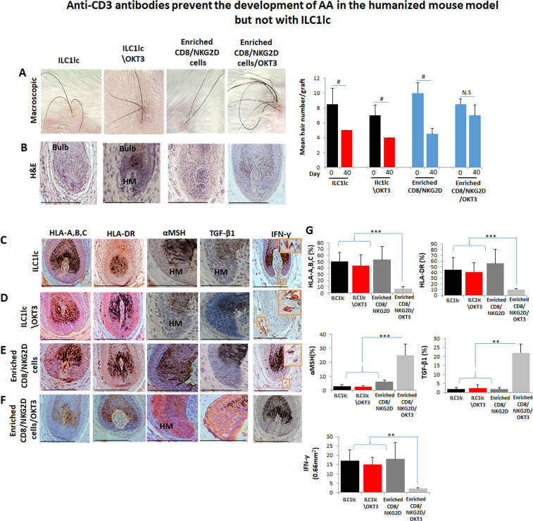
Figure 7—figure supplement 1. Anti-CD3 antibodies prevent the development of alopecia areata (AA) in scalp skin xenotransplants treated with enriched CD8/NKG2D but not with ILC1lc.

Figure 7—figure supplement 1.

(A) Significant hair loss was observed following the injection of ILC1lc, ILC1lc /OKT3, or enriched CD8/NKG2D cells into normal scalp skin on SCID/beige mice, but not in those treated with enriched CD8+/NKG2D+/anti-CD3 (OKT3), N=5 xenotransplants/group from two independent donors. Following Shapiro-Wilk test, Mann-Whitney U test: #p<0.05, ##p<0.01. Hair follicle (HF) dystrophy combined with perifollicular lymphocytic infiltrate around anagen HFs (H&E staining) in xenotransplants treated with (B) ILC1lc, ILC1lc /OKT3, or enriched CD8/NKG2D. Normal HF in anagen in xenotransplant treated with enriched CD8/NKG2D/OKT3. Induction of HLA-A,B,C and of HLA-DR, downregulation of α-MSH and TGF-β1 by the follicular epithelium and increased dermal IFN-γ cells (IHC staining) in xenotransplants treated with (C) ILC1lc and similarly in (D) ILC1lc /OKT3 and (E) enriched CD8/NKG2D treated groups. Reduced expression of HLA-A,B,C, HLA-DR, and upregulation of α-MSH and TGF-β1 combined with decreased number of dermal IFN-γ cells were observed in xenotransplants treated with (F) enriched CD8/NKG2D/OKT3 cells. (G) Quantitative data, N=5–6 xenotransplants/group from two independent donors. 3–4 areas were evaluated per section, and three sections per xenotransplant. Following Shapiro-Wilk test, Student’s t-test: *p<0.05, **p<0.01, ***p<0.001. Scale bar, 50 µm. HM - hair matrix.
Figure 7—figure supplement 1—source data 1. Quantitative data for anti-CD3 antibodies prevent the development of AA in scalp skin xenotransplants treated with enriched CD8/NKG2D but not with ILC1lc.
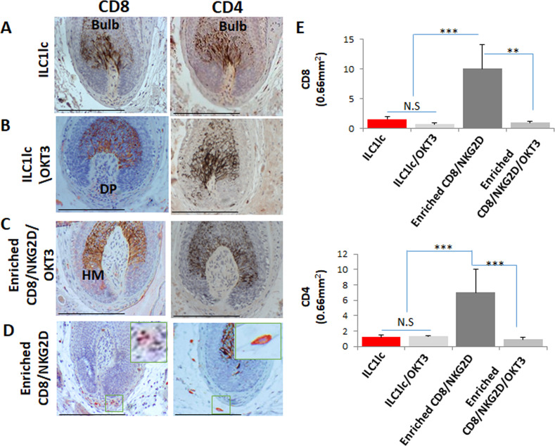
Figure 7—figure supplement 2. Dermal infiltrates of the various treated xenotransplants.

Figure 7—figure supplement 2.

Several CD8 and CD4 in xenotransplants treated with (A) ILC1lc and similarly in xenotransplants treated with (B) ILC1lc /OKT3, and (C) enriched CD8/NKG2D/OKT3 cells versus increased CD8 and CD4 in xenotransplants treated with (D) enriched CD8/NKG2D cells. (E) Quantitative data,N=5–6 xenotransplants/group from two independent donors, 3–four areas were evaluated per section, and three sections per xenotransplant. Following Shapiro-Wilk test, Student’s t-test: *p<0.05, **p<0.01, ***p<0.001. Scale bar, 50 µm. DP - dermal papilla,HM - hair matrix.
Figure 7—figure supplement 2—source data 1. Quantitative data for dermal infiltrates of the various treated xenotransplants.
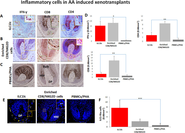
Figure 7—figure supplement 3. Development of alopecia areata (AA) in normal human scalp skin xenotransplants treated with (A) ILC1lc.

Figure 7—figure supplement 3.

Infiltrates of IFN-γ +, CD8, and CD4 cells in group treated with (B) enriched CD8/NKG2D cells. Decreased number of IFN-γ +, CD8, and CD4 cells in the (C) PBMCs/PHA treated one. (D) Quantitative data (E) presence of ILC1lc in transplants injected with ILC1lc compared to the virtual absence of these cells in enriched CD8/NKG2D and PBMCs/PHA treated mice.(F) Quantitative data N=5–9 xenotransplats/group from three donors. 4–5 areas were evaluated per section, and three sections per xenotransplant. Following Shapiro-Wilk test, Student’s t-test: *p<0.05, **p<0.01, ***p<0.001. Scale bar, 50 µm. DP - dermal papilla,HM - hair matrix.
Figure 7—figure supplement 3—source data 1. Quantitative data for development of AA in normal human scalp skin xenotransplants treated with ILC1lc, enriched CD8/NKG2D and PBMCs/PHA.