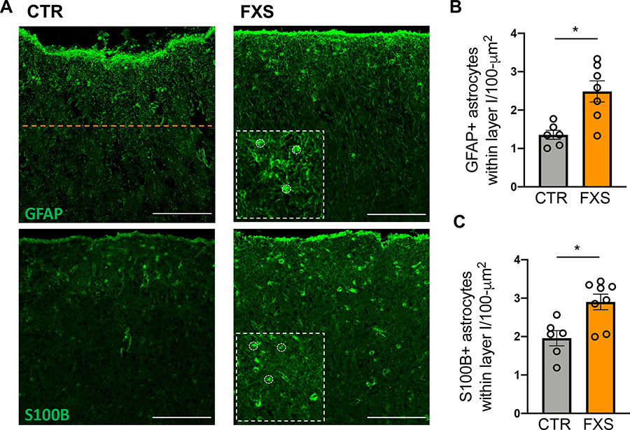
Figure 4. Astrocyte density is higher in postmortem tissue of FXS patients.
A. Representative images of astrocytes in human postmortem cortical sections stained with GFAP and S100B. The orange line indicates the boundary of cortical layer I from the pial surface. Scale bar: 200 μm. Insets: higher magnification with some of the counted astrocytes outlined. B. Density of GFAP+ astrocytes (per 100 μm2) within cortical layer I in postmortem tissue. *P < 0.05, N = 6–8 donors C. Density of S100B+ astrocytes (per 100 μm2) within cortical layer I in postmortem tissue. *P < 0.05, Mean ± SEM.

