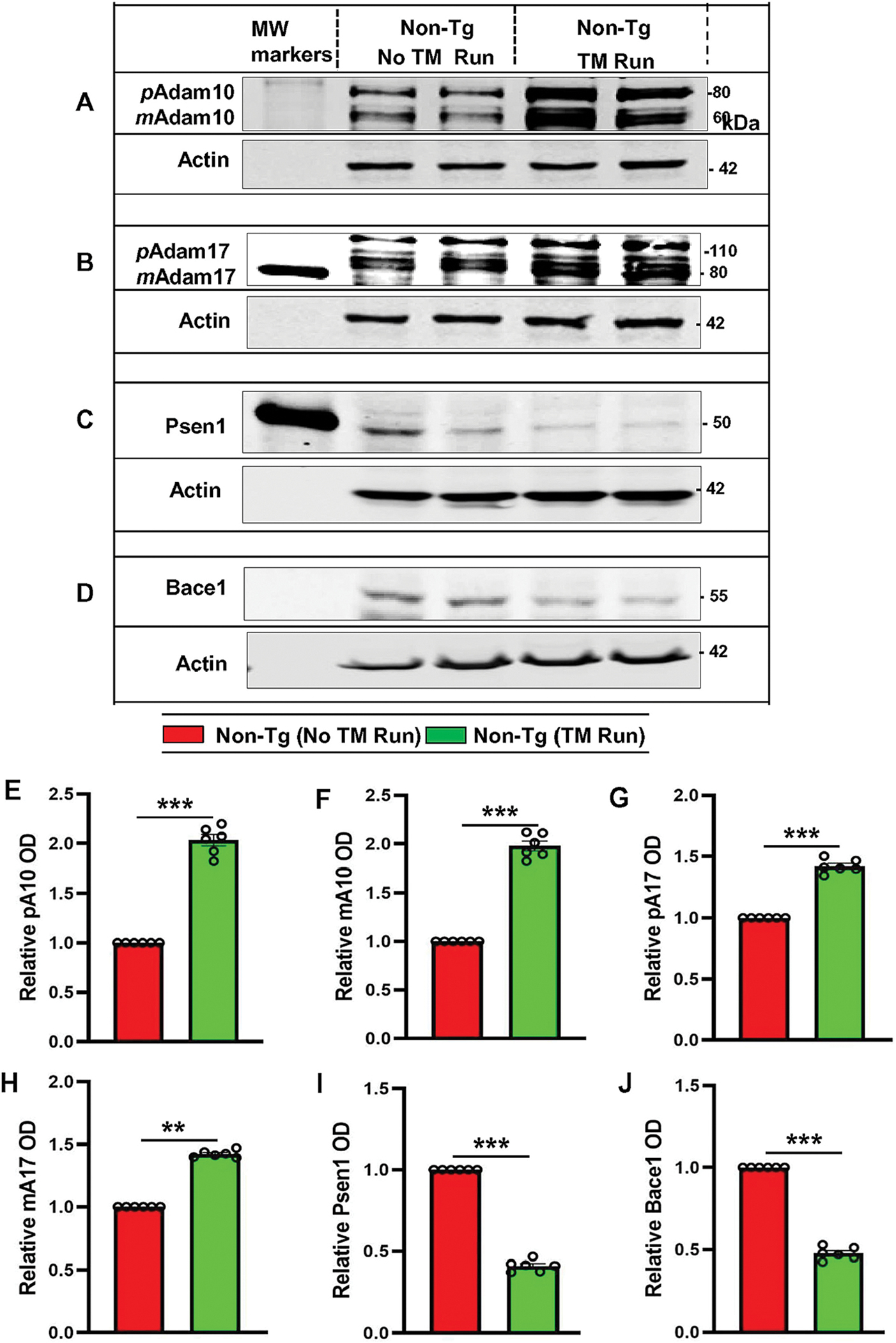
Figure 2. Treadmill exercise results in upregulation of ADAM10, ADAM17, PSEN1, and BACE1 in vivo in the hippocampus of non-Tg mice.

Six-month-old non-Tg mice (n=6/group) were allowed to gently run in the treadmill. After treadmill exercise, mice were sacrificed for monitoring the protein levels – pADAM10, mADAM10, pADAM17, mADAM17, PSEN1 and BACE1 in hippocampal tissue by Western Blot (A-D). Actin was used as the loading control. Bands were scanned and quantified using the NIH Image J software for pADAM10 (E), mADAM10 (F), pADAM17 (G), mADAM17 (H), PSEN1 (I), and BACE1 (J) and the results are represented as relative to non-Tg mice. Results are mean ± SD of six per group. Statistical analysis was conducted by using One-way ANOVA followed by Tukey’s multiple comparison tests. pADAM10 - ***p<0.001 (=0.001498) vs non-Tg mice with exercise; mADAM10 - **p<0.01 (=0.012013) vs non-Tg mice with exercise; pADAM17 – ***p<0.001(=0.000827) vs non-Tg mice with exercise; mADAM17 - **p<0.01(=0.019092) vs non-Tg mice with exercise; PSEN1 - ***p<0.001 (=0.000397) vs non-Tg mice with exercise and BACE1 - ***p<0.001 (=0.009281) vs Non-Tg mice with exercise. Abbreviations: pADAM10 - proADAM10; mADAM10 - matureADAM10; pADAM17 - proADAM17; mADAM17 - matureADAM17; ns – Non-significant.
