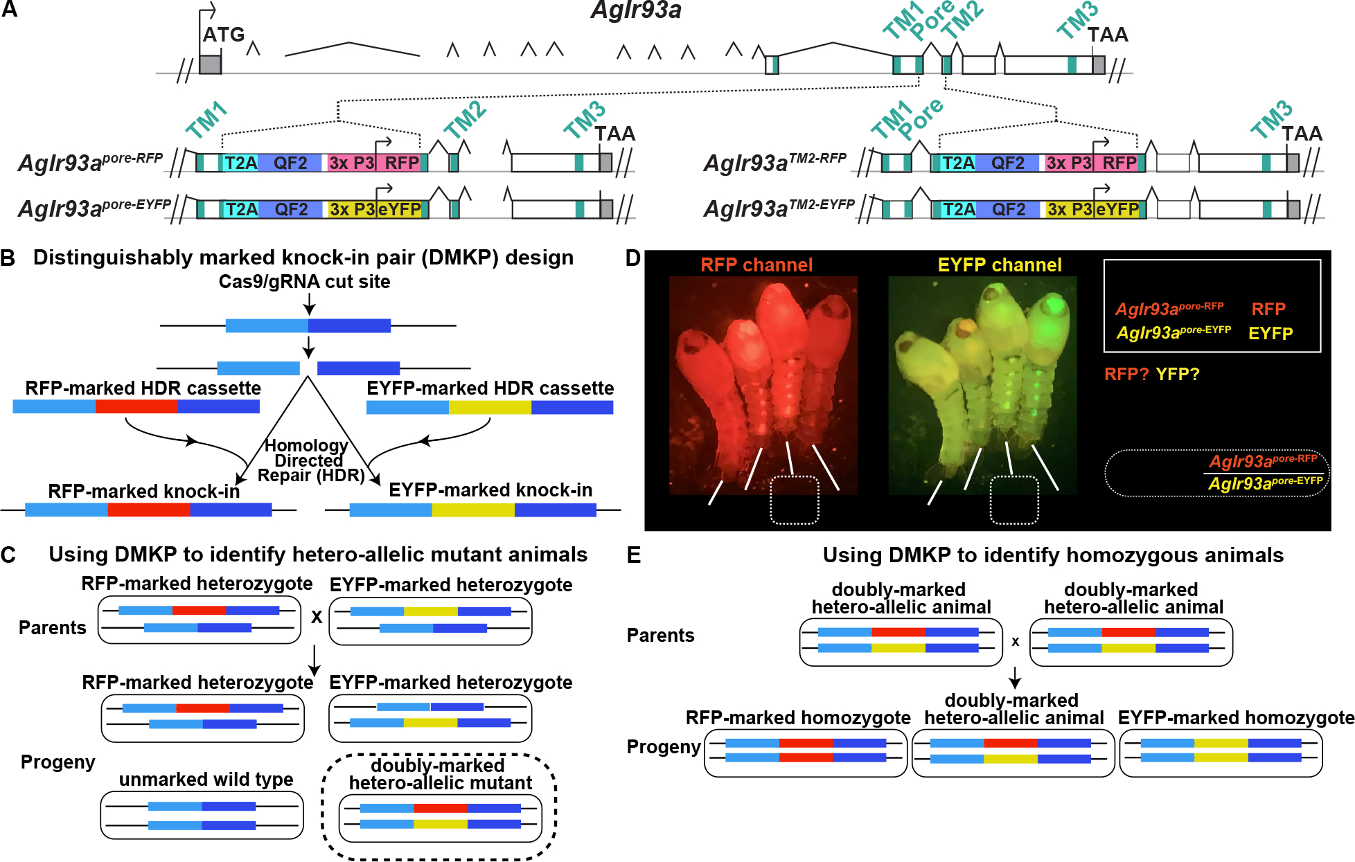
Figure 2: AgIr93a QF2 knock-in animals and the Distinguishably Marked Knock-in Pair (DMKP) strategy.

(A) Top depicts AgIr93a locus organization. Exonic regions encoding the three transmembrane domains and ion pore are denoted in blue-green. 5’ and 3’ UTR are gray. Bottom panels depicts gene structure in AgIr93apore and in AgIr93aTM2 alleles. Each allele was produced by insertion of a knock-in cassette at the location indicated. T2A sequences allow expression of two independent polypeptides from a single mRNA, permitting the expression of the QF2 transcription factor as a separate polypeptide from a transcript initiated at the endogenous AgIr93a start site. The 3xP3 promoter was used to drive visual system and ventral cord expression of the fluorescent transformation marker, which was either RFP or EYFP. (B) Generation of two knock-ins at the same genomic position, identical except that one knock-in is marked with RFP and the other with EYFP. (C) Creating a distinguishably marked hetero-allelic mutant animal using the DMKP strategy. (D) AgIr93apore DMKP pupae. Note that the AgIr93apore-RFP/AgIr93apore-EYFP hetero-allelic mutant pupae express both RFP and EYFP, while heterozygotes and wild type animals do not. Arrowheads highlight RFP and EYFP expression patterns. (E) Generating reliably homozygous knock-in strains by interbreeding hetero-allelic parents using DMKP.
