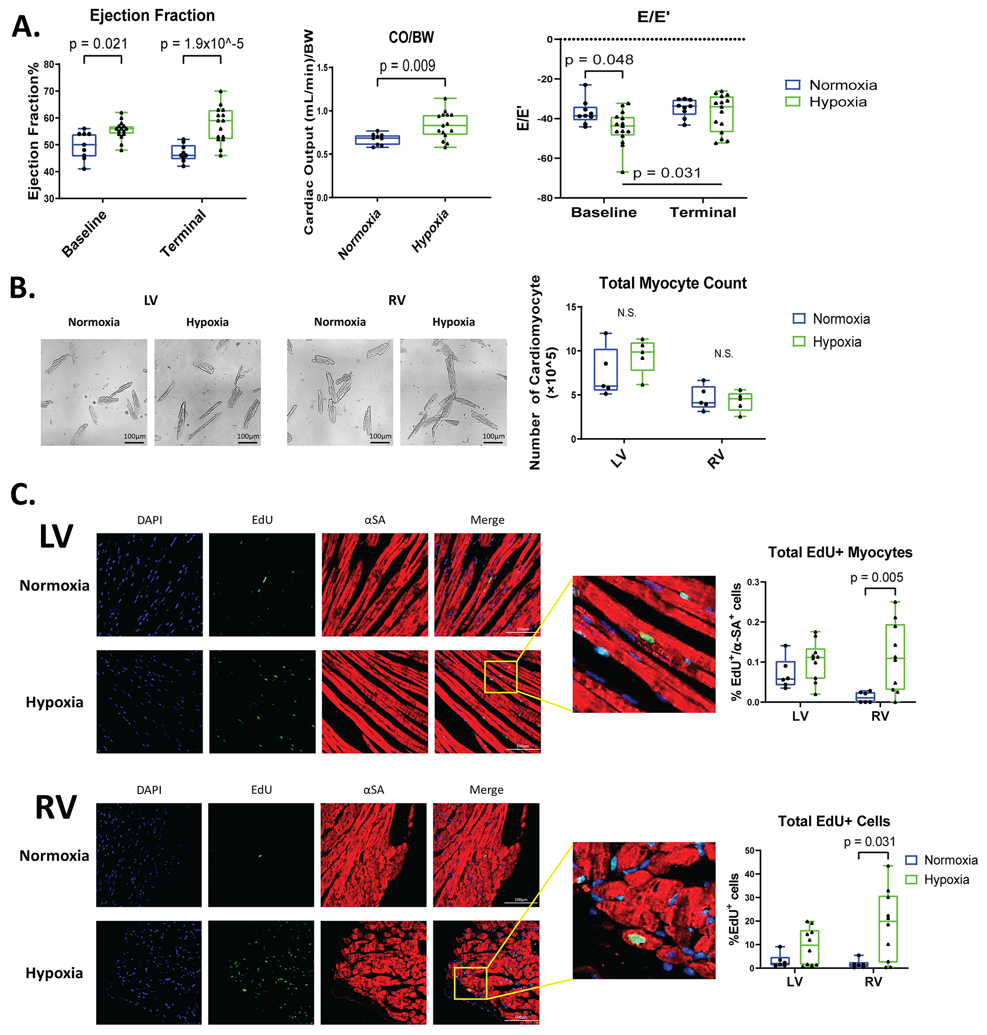
Figure 2. Male mice have normal cardiac function, no changes in the number of LV and RV myocytes, and only RV cardiomyocytes have increased EdU labeling after hypoxia.

A: Ejection fraction (EF), cardiac output relative to body weight (CO/BW), and E/E’ were measured with Echocardiography at baseline and during terminal studies (n=9-15). B: Cardiomyocytes were isolated from the LV and RV of these hearts and the total number of cardiomyocytes were quantified. C: Mouse hearts were stained for alpha-sarcomeric actin (αSA, red, label cardiomyocytes), EdU (green, DNA synthesis), and DAPI (blue, nuclei) to identify myocytes and nonmyocytes undergoing DNA synthesis. Total EdU+ cardiomyocytes and total EdU+ cells were quantified in LV and RV tissue sections (n=6-10). Data are shown as mean±SD.
