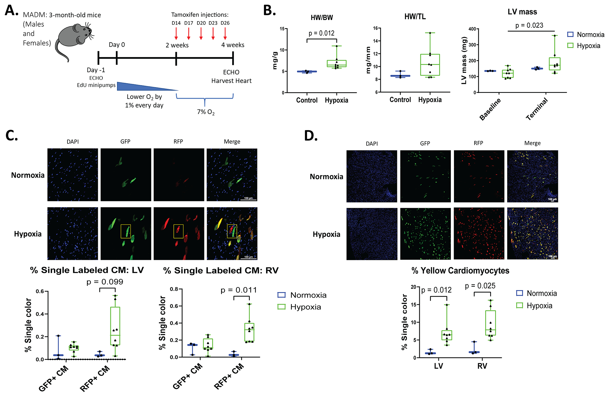
Figure 4. Hypoxia increases the number of labeled cardiac myocytes after tamoxifen injection in MADM mice and identifies an increased number of newly formed myocytes in the RV.

A: Schematic of experimental design in Mosaic analysis with double markers (MADMMyh6-MerCreMer) mice. Five injections of tamoxifen were administered to MADMMyh6-MerCreMer mice every 3 days (D) on day 14, 17, 20, 23, and 26. B: HW/BW and HW/TL was determined during terminal studies. Echocardiography was performed at baseline and during terminal studies to measure LV mass. C and D: Following tamoxifen injections, myocytes that completed cell division were single-labeled GFP+ or RFP+ only (“twin spots” highlighted by the yellow box in D). Hypoxia increased the number of affected cardiac myocytes that were labeled yellow (GFP and RFP double positive) in the LV and RV. Quantification of green (GFP+), red (RFP+), and yellow myocytes in male and female mice (normoxia n=3, hypoxia n=8). Data represented as mean±SD.
