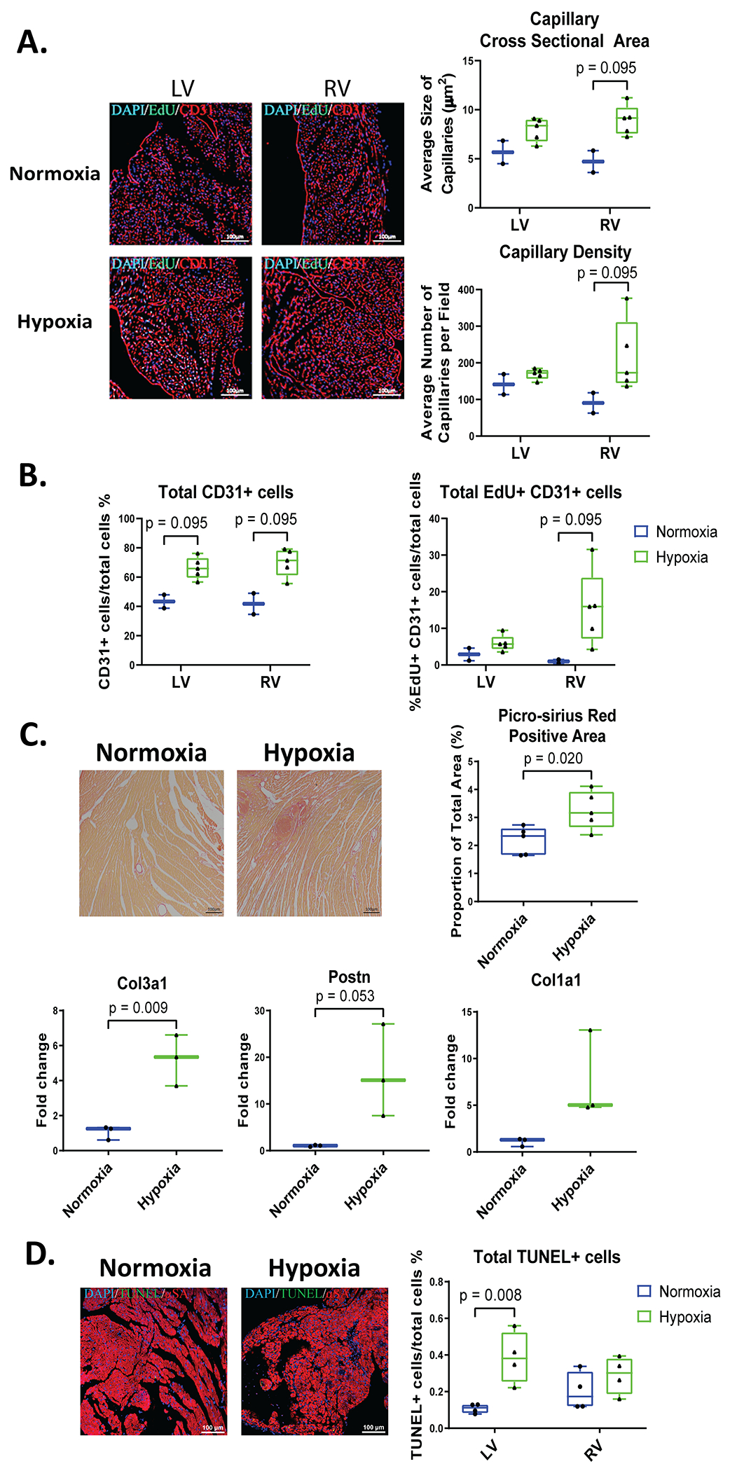
Figure 5. Systemic Hypoxemia stimulates cardiac remodeling with fibrosis and non-myocyte cell proliferation.

Adult hearts were stained for CD31 (red, labels endothelial cells), EdU (green), and DAPI (blue). A: Capillary density and size was measured in LV and RV sections of the heart. B: Total CD31+ cells and total EdU+ CD31+ cells were quantified. C: Fibrosis was analyzed and quantified by Picro Sirius Red staining and gene expression level of collagen 3a1 (col3a1), collagen 1a1 (col1a1), and Periostin (Postn) by RT-PCR. D: Heart sections were stained for αSA (red, labels cardiomyocytes), TUNEL (green, apoptotic cells), and DAPI (blue, nuclei) to label total apoptotic (TUNEL+) cells (n=4-5 mice). Data represented as mean±SD.
