
Extended Data Fig. 3. Structures of the capsomers in IP6-induced in vitro CLPs are similar to their corresponding structures in situ.
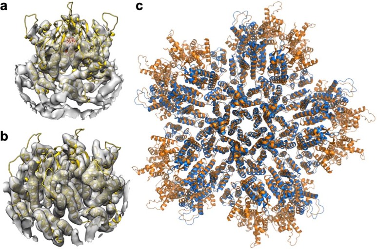
(a) Docking of our real-space-refined CA pentamer model (yellow ribbons, with IP6 in sticks) into EMD-3466 (gray), the first reported in situ structure of the HIV-1 CA pentamer at 8.8 Å resolution7. (b) Docking of our real-space-refined CA hexamer model (yellow ribbons) into EMD-3465 (gray), an in situ structure of the HIV-1 CA hexamer at 6.8 Å resolution7. (c) Superposition of the final PDB model of the declination after real space refinement into our 3.4 Å map (orange) with PDB 5mcy (ref. 7) (blue).