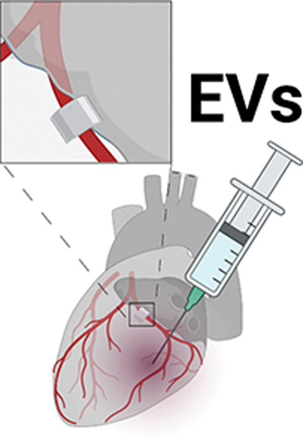
Figure 1: Graphical abstract.
Swine at age 6 weeks were given four weeks of high fat and high cholesterol diet to induce metabolic syndrome. An ameroid constrictor was placed on the proximal left circumflex artery to induce chronic myocardial ischemia. Two weeks later, animals received intramyocardial injection of either saline vehicle (“control,” n=6) or extracellular vesicles (high fat diet with myocardial EV injection group “HVM,” n=8). Five weeks later myocardial tissue was harvested for analysis. Ischemic myocardium in the HVM group had decreased expression of anti-angiogenic signaling markers angiostatin and endostatin. Angiostatin expression localized the coronary vasculature as determined by α-smooth muscle actin (α-SMA) staining. Among EV-treated pigs, angiostatin expression was inversely related to blood flow to ischemic myocardium during pacing. EV = extracellular vesicle. In boxplots, upper and lower borders of box represent upper and lower quartiles, middle horizontal line represents median, upper and lower whiskers represent maximum and minimum values of non-outliers. Created with BioRender.com.

