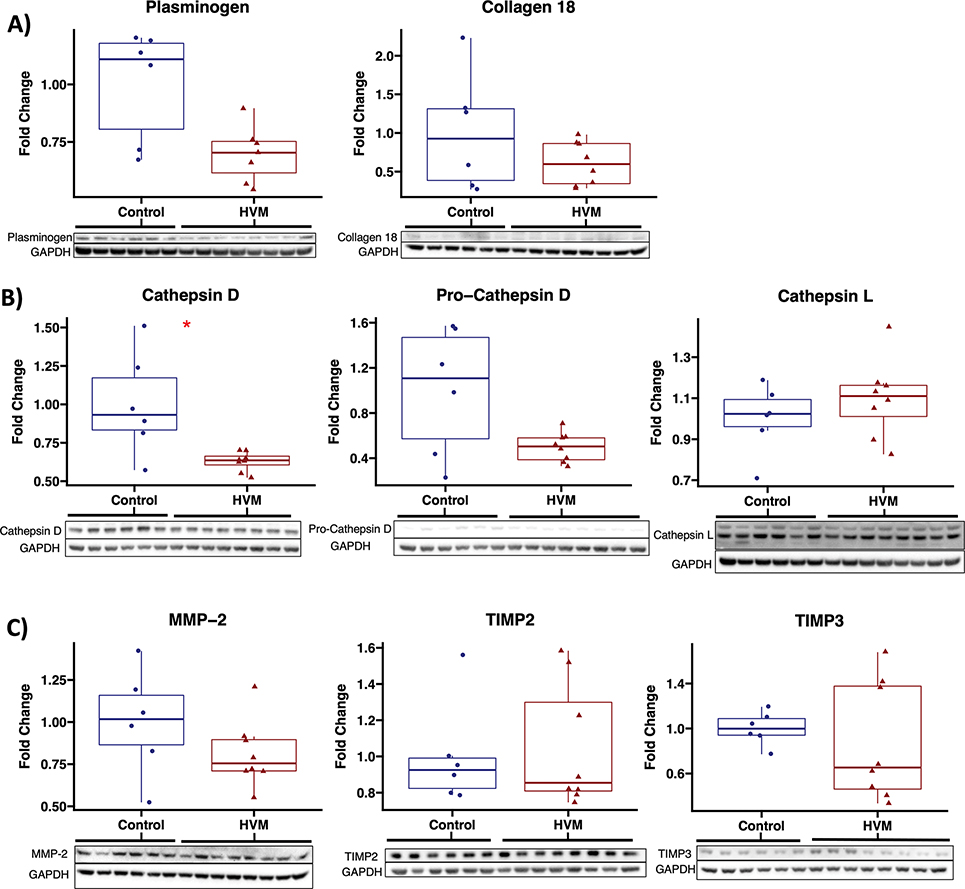
Figure 5: Among proteins involved in production of angiostatin and endostatin, cathepsin D is significantly decreased with EV therapy.
Immunoblot results are shown for A) precursors to angiostatin and endostatin, B) cathepsins D and L which cleave precursors to generate angiostatin and endostatin, and C) matrix metalloproteinase-2 (MMP-2) and tissue inhibitor of metalloproteinases (TIMP) 2 and 3 which regulate generation of angiostatin and endostatin. HVM, high fat diet swine that received intramyocardial extracellular vesicles (n=8), control, high fat diet swine that received saline vehicle (n=6). Upper and lower borders of box represent upper and lower quartiles, middle horizontal line represents median, upper and lower whiskers represent maximum and minimum values of non-outliers. *p<0.05 as determined by Wilcoxon rank-sum test.

