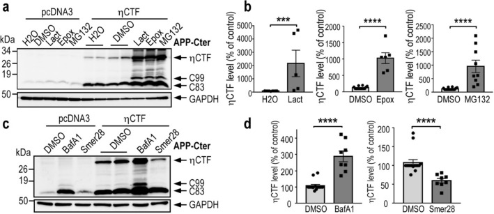
Fig. 2.
ηCTF fragment is degraded by both proteasome and autophagic pathways. a–d SH-SY5Y cells were transiently transfected with ηCTF or pcDNA3 vectors and treated for 24 h with proteasome inhibitors (a, b, lactacystine (Lact, 5 µM), epoxomicin (Epox, 1 µM), MG132 (5 µM)) or with bafilomycin A1 (BafA1, 100 nM) or Smer28 (50 µM) that blocks or activates autophagy respectively (c, d) then analyzed by western blot using APP-Cter antibody. Histograms in b, d correspond to the quantification of ηCTF immunoreactivity obtained in a, c and are expressed as percent of controls (H2O or DMSO-treated cells) taken as 100. Bars are the means ± SEM of 5–9 independent determinations. ****p < 0.0001 according to Mann–Whitney test. All full gels are provided in Sup Fig. 5

