Fig. 7.
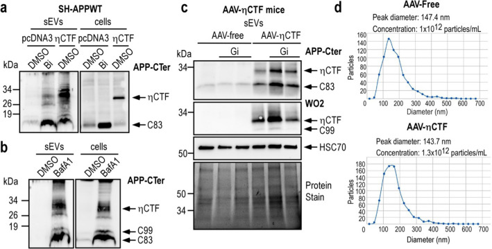
ηCTF fragment is detected in sEVs purified from cells and mouse brains. a–b SH-APPWT cells were transiently transfected with ηCTF or empty pcDNA3 vector and treated for 24 h with β-secretase inhibitor (a, Bi, 30 µM), or bafilomycin A1 (b, BafA1, 100 nM). Cell lysates and sEVs were purified from culture media as described in methods and analyzed by western blot using APP-Cter antibody. c sEVs were purified from brain homogenates of 3-month-old AAV-free and AAV-ηCTF mice in the presence or not of the α-secretase inhibitor (Gi:10 µM) and analyzed by western blot using APP-Cter antibody. HSC70 is used as an exosomal marker. Whole loaded proteins were stained by photoactivation using Bio-Rad prestain method (Protein Stain) as loading control. All full gels are provided in Sup Fig. 5. d Concentration and particles size of each brain mouse exosomal purified samples were analyzed in ZetaView instrument (Particle-Metrix) before loading on gels

