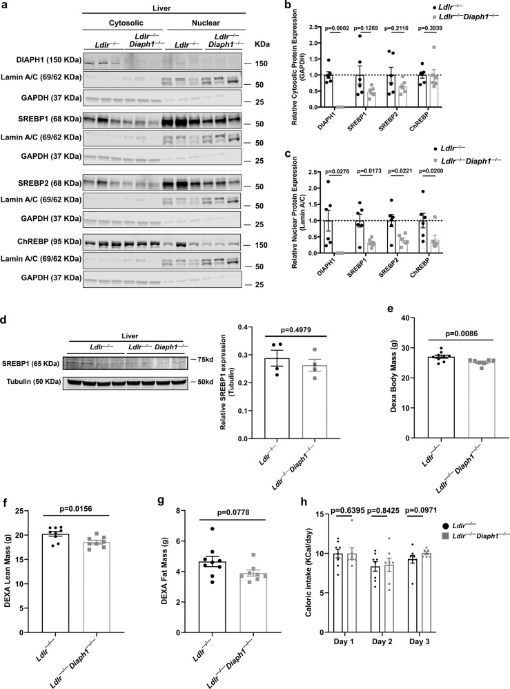
Fig. 5. Deletion of Diaph1 in Ldlr−/− mice reduces nuclear content of SREBP1, SREBP2 and ChREBP in liver.
Ldlr −/− and Ldlr−/− Diaph1−/− male mice were fed WD for 16 weeks. a Representative Western Blots for the detection of cytosolic and nuclear DIAPH1, SREBP1, SREBP2 and ChREBP performed on liver fractions isolated from the indicated mice. b Quantification of cytosolic DIAPH1, SREBP1, SREBP2 and CHREBP, relative to GAPDH. c Quantification of nuclear DIAPH1, SREBP1, SREBP2 and ChREBP, relative to Lamin A/C. d Representative Western blot and quantification of total SREBP1 normalized to tubulin in total liver of the indicated mice. e–g DEXA scans were performed for determination of body mass (e), lean mass (f), and fat mass (g). h Caloric intake was determined over 3 consecutive days. i mRNA expression of the gene encoding RAGE (Ager) was determined in the livers of the indicated male mice after 16 weeks WD. The mean ± SEM is reported. The number of independent mice/group is indicated in the figure as individual data points. Statistical analyses regarding testing for the normality of data followed by appropriate statistical analyses were described in Materials and Methods. P-values were determined by unpaired T-test or Wilcoxon rank-sum test depending on if the data passed the Shapiro-Wilk normality test.

