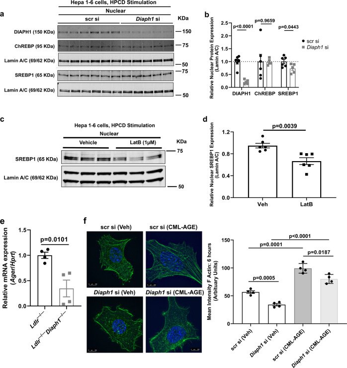
Fig. 8. RAGE, DIAPH1, actin organization and SREBP1.
a Western blots for the detection of nuclear DIAPH1, ChREBP and SREBP1 performed on mouse Hepa 1-6 cells after Diaph1 or scramble control siRNA knockdown and 30-min sterol depletion with 1% 2-hydroxypropyl-β-cyclodextrin (HPCD). b Quantification of nuclear DIAPH1, SREBP1, and ChREBP, relative to Lamin A/C. c Western blots for the detection of nuclear SREBP1 performed on mouse Hepa 1-6 cells after 30 min pre-treatment with latrunculin B (LatB; 1 µm) followed by the addition for 30 min of sterol depletion with 1% HPCD. d Quantification of nuclear SREBP1 relative to Lamin A/C. e mRNA expression of the gene encoding RAGE (Ager) was determined in the livers of the indicated male mice after 16 weeks WD. f Hepa 1-6 cells bearing scramble control or Diaph1 siRNA silencing were treated with RAGE ligand CML-AGE (100 µg/ml) or vehicle for 6 h followed by quantification of the mean intensity of F-actin (phalloidin). Scale bar: 250 µm. The mean ± SEM is reported. The number of independent biological/independent replicates is indicated in the figure as individual data points. Statistical analyses regarding testing for the normality of data followed by appropriate statistical analyses were described in Materials and Methods. P-values were determined by unpaired T-test or Wilcoxon rank-sum test depending if data passed the Shapiro-Wilk normality test.

