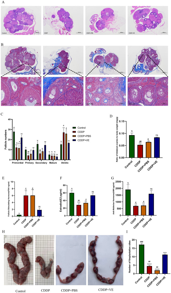
Figure 2.
Effects of CDDP and ferroptosis inhibitor Vitamin E on ovarian function and fertility in rats. (A, C) HE staining of the ovarian tissue structure in each group and the statistical results of the number of follicles in different developmental stages. (B, D) Masson trichrome staining of the ovarian tissue in each group observed under a microscope. (E, F, G) Serum FSH, E2 and AMH levels in each group. (H) Gross observation of uterine litter size of rats in each group. (I) Summary of embryo numbers at implantation sites of each group. The score of stained Masson trichrome Staining was quantitated using ImageJ software. Data are expressed as the means ± SEM. ns, * versus control, *P < 0.05, **P < 0.01, ***P < 0.001 and ****P < 0.0001, ns indicates no statistical significance. N, # versus CDDP + VE, # P < 0.05, ## P < 0.01, ###P < 0.001 and #### P < 0.0001, N indicates no statistical significance.

