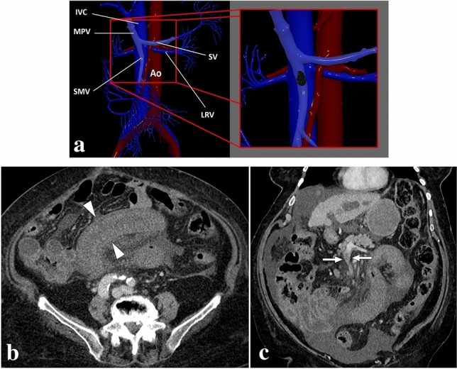
Fig. 13.

a 3D illustration of SMV and its branches for a quick review. The magnified view represents a thrombus in the main SMV branch (IVC inferior vena cava, HPV hepatic portal vein, SV splenic vein, Ao abdominal aorta, SMV superior mesenteric vein, LRV left renal vein). b An 83-year-old male patient with known hepatocellular carcinoma presented with acute onset diffuse abdominal pain. Axial plane postcontrast CT showed symmetrical wall thickening in a long segment of small bowel loops (arrowheads) accompanied by mesenteric fat stranding. c Coronal plane CT image demonstrated thrombosed mesenteric segmental vein (arrows) draining the affected small bowel loops. Imaging findings were consistent with segmental veno-occlusive mesenteric ischemia, and the patient was treated accordingly
