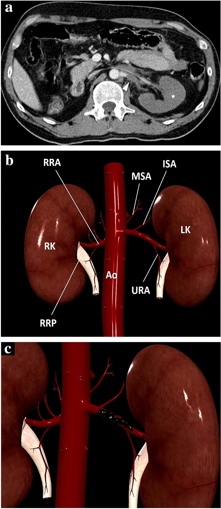
Fig. 15.

a A 39-year-old male patient with a known left solitary kidney was brought to the ER after high-energy trauma. Axial plane post-contrast abdominal CT image showed a thrombosed left renal artery (arrowhead) and complete devascularization of the left renal parenchyma with no apparent parenchymal enhancement (asterisk). An arterial graft was surgically placed between the abdominal aorta and left renal hilus to establish renal vascularization. b 3D illustration indicates renal arteries and some important major branches for a quick review. MSA middle suprarenal artery, ISA inferior suprarenal artery, URA ureteric branches of the renal artery, Ao Abdominal aorta, RRA right renal artery, RK right kidney, LK left kidney, RRP right renal pelvis. c Representation of thrombus in the lumen of the left renal artery
