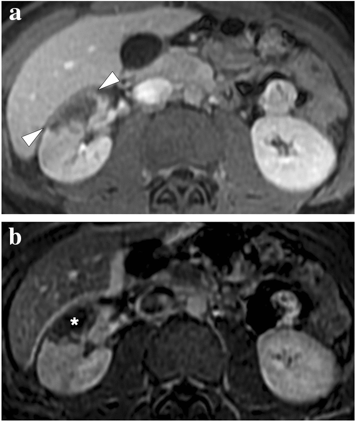
Fig. 16.

A 19-year-old male patient with known hereditary thrombophilia presented with recent onset right flank pain. An initial US examination was unremarkable except for mild heterogeneity, without any obvious mass lesion, in the interpolar region of the right kidney (not shown). a Axial plane T1-weighted dynamic postcontrast abdominal MR image showed a triangular-shaped hypointense, non-enhancing area (arrowheads) in the right kidney, which was found to be suggestive of renal infarction. b Axial plane subtraction image confirmed the absence of focal contrast enhancement (asterisk)
