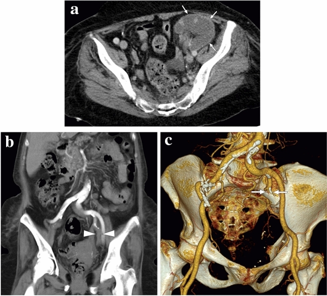
Fig. 17.

A 65-year-old female with a history of renal transplantation nearly 20 years ago presented with severe, acute left lower quadrant pain. a Axial plane postcontrast CT showed complete devascularization of graft kidney with cortical rim sign (arrows). b and c Coronal plane reformatted CT angiography (b) and 3D-VRT images (c), respectively, demonstrated the acute thrombotic occlusion (arrowheads) and the remaining stump of the renal artery (arrows) anastomosed to the left internal iliac artery
