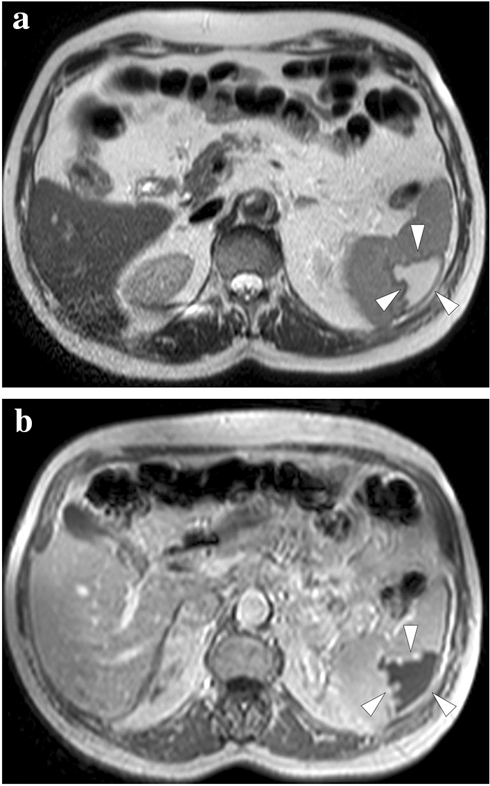
Fig. 20.

A 63-year-old male with previously diagnosed splenic infarction due to atrial fibrillation in an outside institution. a and b Axial plane T2 and post-contrast T1-weighted MR images, respectively, showed peripherally located, triangular-shaped, well-demarcated, T2-hyperintense, and T1-hypointense nonenhancing parenchymal area (arrowheads). These findings were found to represent cystic-liquefying changes in the subacute phase
