Fig. 21.

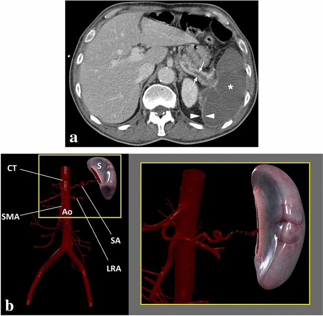
A 68-year-old male patient with known lung cancer and polycythemia vera presented with acute onset severe left upper quadrant pain. a Axial plane postcontrast abdominal CT showed thrombosed splenic artery (arrows) and global splenic infarction (asterisk). Also, note was made of enhancing splenic capsule (arrowheads). b 3D illustration shows the splenic artery and some major branches of the abdominal aorta (S spleen, CT celiac trunk, Ao abdominal aorta, SMA superior mesenteric artery, LRA left renal artery, SA splenic artery). The magnified view demonstrates the thrombosis of the splenic artery
