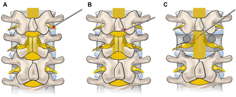
Figure 8.
Schematic representation of transgrade DRG lead placement. (A) depicts the initial position where the Tuohy needle enters the skin. (B) depicts the Tuohy needle contacting the lamina prior to walking off and entering the epidural space. The red cross indicates graphic target for Tuohy needle. (C) depicts the Touhy needle within the epidural space with the introducer sheath and lead positioned along the superior aspect of the contralsteral foramen, one level below. Reprinted with permission from Chapman KB, Ramsook RR, Groenen PS, Vissers KC, van Helmond N. Lumbar transgrade dorsal root ganglion stimulation lead placement in patients with post-surgical anatomical changes: a technical note. Pain Pract. 2020;20(4):399–404.© 2019 World Institute of Pain.179

