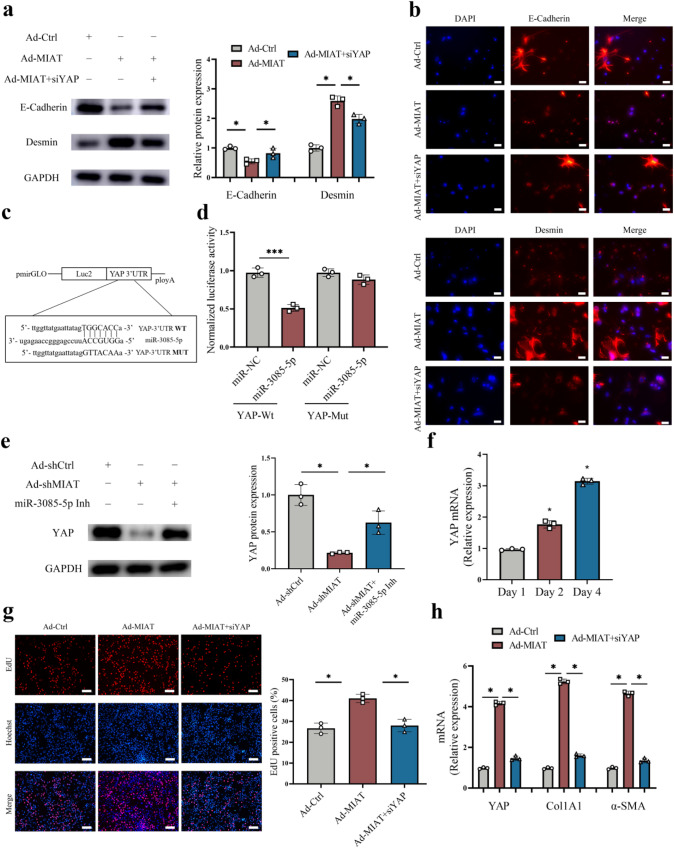
Fig. 6. Loss of MIAT inhibits Hippo/EMT signaling pathway and HSC activation via YAP.
a Protein expressions of E-cadherin and Desmin in HSCs transfected with Ad-Ctrl, Ad-MIAT or Ad-MIAT plus siYAP (n = 3 per group). b Immunofluorescence staining for E-cadherin and Desmin in HSCs transfected with Ad-Ctrl, Ad-MIAT or Ad-MIAT plus siYAP. Scale bar, 20 μm. c Binding sites of miR-3085-5p with respect to YAP. d Luciferase assays of pmirGLO-YAP-Wt or pmirGLO-YAP-Mut in HEK-293T cells with miR-NC or miR-3085-5p (n = 3 per group). e Protein expression of YAP in HSCs transfected with Ad-shCtrl, Ad-shMIAT or Ad-shMIAT plus miR-3085-5p inhibitor (n = 3 per group). f YAP expression in primary HSCs at day 1, 2 and 4 (n = 3 per group). g Cell proliferation in HSCs transfected with Ad-Ctrl, Ad-MIAT or Ad-MIAT plus siYAP (n = 3 per group). Scale bar, 50 μm. h YAP, Col1A1 and α-SMA expression in HSCs transfected with Ad-Ctrl, Ad-MIAT or Ad-MIAT plus siYAP (n = 3 per group). Each value is the mean ± SD of three independent experiments. *p < 0.05.

