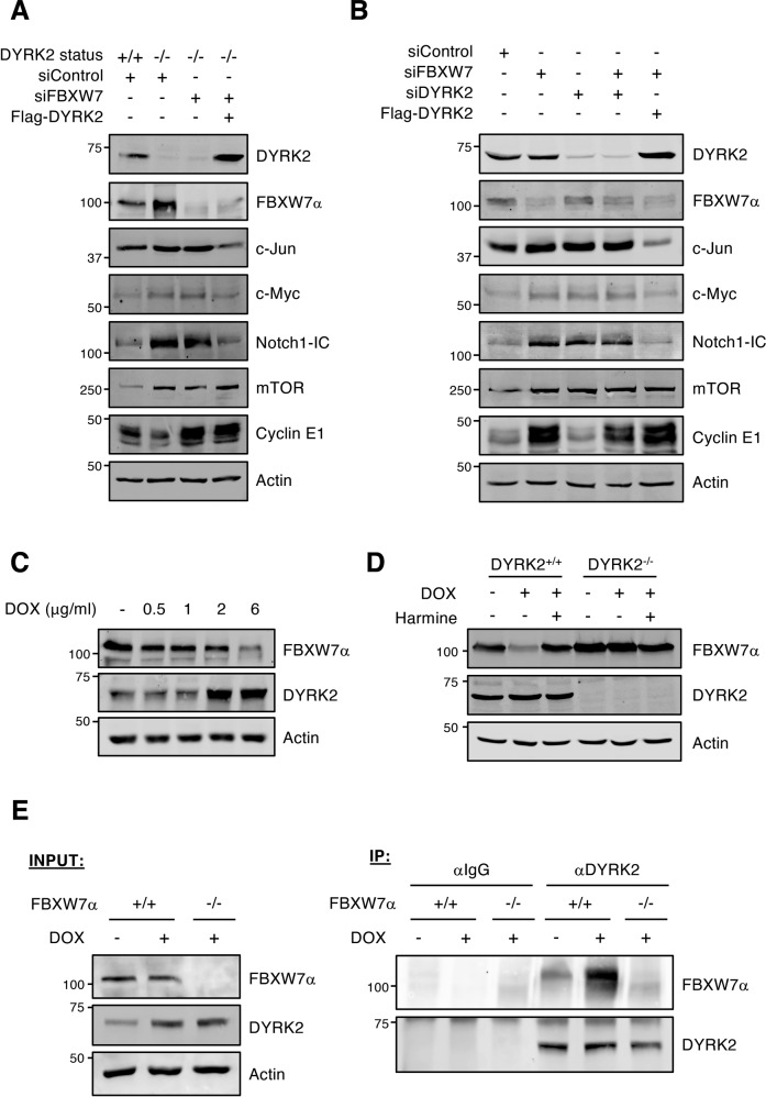
Fig. 6. DYRK2 modulates FBXW7 activity.
A HEK-293T WT (+/+) or DYRK2-KO (−/−) cells were transfected with the indicated siRNAs or plasmids, and the indicated endogenous proteins were analyzed by WB. B HEK-293T cells were transfected with the indicated siRNAs or plasmids, and the endogenous levels of the specified proteins were analyzed by WB. C HEK-293T cells were treated with increasing concentrations of DOX for 12 h, and endogenous FBXW7α and DYRK2 analyzed by WB. D HEK-293T cells and derived DYRK2-KO cells were treated with DOX (2 μg/ml) or vehicle for 12 h in the presence or absence of Harmine (5 μM), and endogenous DYRK2 and FBXW7α analyzed by WB. E HCT116 cells WT (+/+) and knockout (−/−) for FBXW7 were treated with DOX (2 μg/ml) or with vehicle for 12 h. MG-132 (10 μM) was included to stabilized FBXW7. Control and treated extracts (INPUT) were immunoprecipitated with a DYRK2 antibody or control IgGs and the presence of DYRK2 and FBXW7 in the immunoprecipitates analyzed by WB (IP). Note: a representative experiment is shown in each panel of 3–4 performed.

