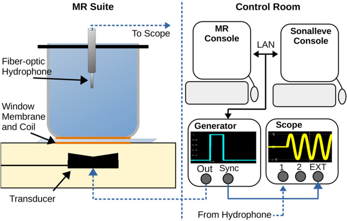
Figure 2.
(Left) The MRI suite contains the Sonalleve treatment bed, ultrasound transducer, quality control phantom, and needle hydrophone. The hollowed phantom is filled with deionized, degassed water and acoustically coupled to the window membrane. (Right) The planning images are sent to the Sonalleve console where a treatment cell was placed 6 cm above the membrane. The external trigger function is used to set the trigger approximately 90 µs after the waveform generator burst to avoid measuring electrical coupling of the incident pulse. Dashed lines indicate wiring through separate rooms.

